Research says fasting while pregnant affects baby’s growth rate, increases risk of adult-onset disorders
07/18/2019 / By Evangelyn Rodriguez
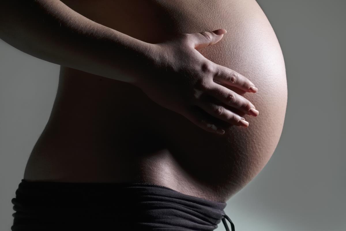
Intrauterine growth restriction (IUGR) is an irreversible complication of pregnancy that results in poor fetal growth. IUGR is caused by maternal food restriction (FR) or malnutrition, but the mechanisms underlying this condition is not fully understood. In a recent study published in the journal Nutrition Research, a team from the David Geffen School of Medicine at the University of California, Los Angeles investigated the molecular pathways involved in the development of IUGR. They evaluated key cytokines like interleukin-10 (IL-10), cellular processes related to immunity, and the resulting IUGR phenotype and placental histopathology using a mouse model.
IUGR and its possible causes
IUGR is a condition in which a fetus suffers from poor or restricted growth. Babies with IUGR are too small for gestational age and may be born prematurely (before 37 weeks). IUGR is said to affect up to 10 percent of pregnancies and often results in short- or long-term sequelae for the offsprings.
IUGR can occur when the mother contracts an infection or the fetus is unable to receive the necessary nutrients for proper growth and development. This is why maternal nutrition and diet are very important considerations during pregnancy. Maternal FR places the offspring at great risk of short-term morbidity or adult-onset cardiovascular and metabolic disorders. (Related: Pregnant women who don’t get enough vitamin D risk having children with schizophrenia.)
Some maternal factors involved in the development of IUGR include:
- Hypertension
- Heart or respiratory disease
- Advanced diabetes
- Cigarette smoking
- Chronic kidney disease
On the other hand, the fetal environment can also influence IUGR development. Some uterus- or placenta-related factors that can cause IUGR are:
- Infection of tissues in the uterus
- Placental abruption
- Placental previa or low attachment in the uterus
- Decreased blood flow in the uterus and placenta
Maternal food restriction decreases anti-inflammatory interleukin-10 expression and affects immunologic pathways in the placenta
The placenta plays an important role in the development or prevention of IUGR. Nutrients obtained by the mother from food is passed to her offspring via blood through this organ. When the mother changes her diet, the placenta is able to sense this and responds to the change in a way that directly affects the transport of nutrients to the fetus. In IUGR, the placenta responds to longstanding nutrient and oxygen insufficiency by exhibiting a certain phenotype. This includes hypovascularity or insufficient blood vessels, fibrin deposition, and obstruction of blood supply in many areas. On the other hand, the maintenance of the placental structure relies on immunologic pathways that allow maternal-fetal immune tolerance.
Previous studies suggested that changes in immune tolerance mediate complications in pregnancy such as IUGR. Interleukin-10 (IL-10), an anti-inflammatory cytokine, is said to be also altered in IUGR. IL-1o is one of the key regulators that makes maternal-fetal immune tolerance in the placenta possible.
In the study, the researchers hypothesized that maternal FR during pregnancy would decrease the overall immunotolerant environment in the placenta, leading to placental insufficiency, increased cellular stress and death, and the development of IUGR. They tested their hypothesis by subjecting pregnant mice to mild and moderate FR from gestational day 10 to 19. They collected their placentas and embryos at gestational day 19 for further evaluation.
To identify immunologic pathways affected in IUGR-associated placentas, the researchers used RNA sequencing. They also validated the messenger RNA expression changes of genes important for cellular integrity. In addition, they evaluated histopathological changes in vascular and trophoblastic structures as well as protein expression changes in autophagy, endoplasmic reticulum (ER) stress, and apoptosis. Trophoblasts are the placental cells responsible for providing nutrients to the embryo. In IUGR, both vascular and trophoblast structures are downregulated.
The researchers identified several differentially expressed genes in FR, including a considerable subset that regulates immune tolerance, inflammation, and cellular integrity. They also found that IL-10 and some autophagic genes are suppressed in FR-induced IUGR. While they were right about IL-10 inhibition, FR did not increase autophagy and ER stress. The researchers believed that the placenta prioritizes fetal growth even in the face of adverse maternal environment. They also believed that the mechanisms underlying this function are immunologically mediated.
The researchers concluded that maternal food restriction induces the development of fetal growth restriction by inhibiting the anti-inflammatory protein IL-10 and the suppression of placental autophagic and ER stress responses.
Visit WomensHealth.news for more stories and studies on the factors that affect proper fetal growth.
Sources include:
Tagged Under: anti-inflammatory, autophagy, cytokines, fasting, fetal growth, fetal health, fetus, food restriction, gestation, immune system, immunotolerance, intrauterine growth restriction, maternal diet, maternal food restriction, maternal nutrition, placenta, pregnancy, pregnant mothers, research, trophoblast
RECENT NEWS & ARTICLES
ImmuneSystem.News is a fact-based public education website published by Immune System News Features, LLC.
All content copyright © 2018 by Immune System News Features, LLC.
Contact Us with Tips or Corrections
All trademarks, registered trademarks and servicemarks mentioned on this site are the property of their respective owners.


